this post was submitted on 13 Jul 2023
9 points (100.0% liked)

Radiology

598 readers
1 users here now

A community for all things related to medical imaging!

RULES:

-1. Please follow the Lemmy.World Server Rules.

-1A. Please be civil.

-1B. Please be respectful when discussing medical cases. While we do not wish to impose a somber tone upon this community, please remember that there are real patients behind the images.

-2. No patient-identifiable information. There is zero tolerance for breaching patient confidentiality laws. De-identified information is allowed under HIPAA, the US patient confidentiality law. Consent is not required when posting de-identified information.

-3. No requests for medical advice or second opinions. Please go to your physician/provider for assistance. Online strangers will never know your clinical history as well as your actual healthcare team, nor will the images posted here be of sufficient quality or completeness for diagnostic interpretation. Any content or discussions in this Community should be considered for educational purposes only, and their accuracy or quality with regards to standard of care cannot be guaranteed.

-4. No spam or advertising. Products or companies that are mentioned as a natural course of discussion are allowed.

-5. Please do not spread misinformation.

-6. Moderators have final say in their decisions. Please, no rules lawyering.

Posts:

Only moderators may post in this community pending Lemmy's implementation of a moderator-approval process. Until that happens, you may post general comments or questions in the stickied megathread. You may also request the moderators to post on your behalf via DM, pending time and availability.

founded 2 years ago
MODERATORS
 

59 year old with no past medical history, now with new dyspnea on exertion.

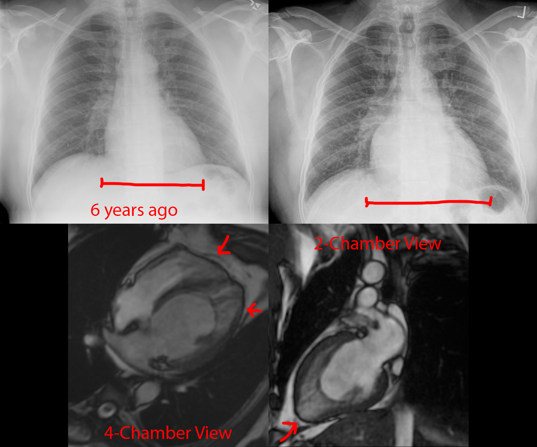
Top: Chest radiographs show development of cardiomegaly since 6 years prior. The pulmonary vascular markings are also more prominent than before, suggesting pulmonary congestion.

Bottom: MRI Cine FIESTA sequences in the 4- and 2-chamber views at end-diastole. There are prominent trabeculations towards the cardiac apex at both the left and right ventricles. In particular, there is an outer layer of dense myocardium and a thicker inner layer of trabeculated myocardium.

Final Diagnosis: Cardiac noncompaction.

Comparison Case: Meth-Induced Cardiomyopathy.

Cardiac noncompaction is a genetic disorder leading to failure of the myocardium to form "compact" walls. It is extremely rare. The presentation depends on how bad the noncompaction is, so can be asymptomatic to heart failure, arrhythmias, and thromboembolism.

no comments (yet)
sorted by: hot top controversial new old
there doesn't seem to be anything here