this post was submitted on 20 Nov 2024
25 points (93.1% liked)

Forage Fellows ๐Ÿ„๐ŸŒฑ

436 readers
6 users here now

Welcome to all things foraging! A new foraging community, where we come together to explore the bountiful wonders of the natural world and share our knowledge of gathering wild goods! ๐ŸŒฑ๐Ÿ“๐Ÿซ

founded 1 year ago
MODERATORS
 

View the spoiler for my guess at what I think it might be, but please first come to your own conclusion before looking at mine โ€” I don't want to bias your guess.

My guessPsilocybe cyanescens


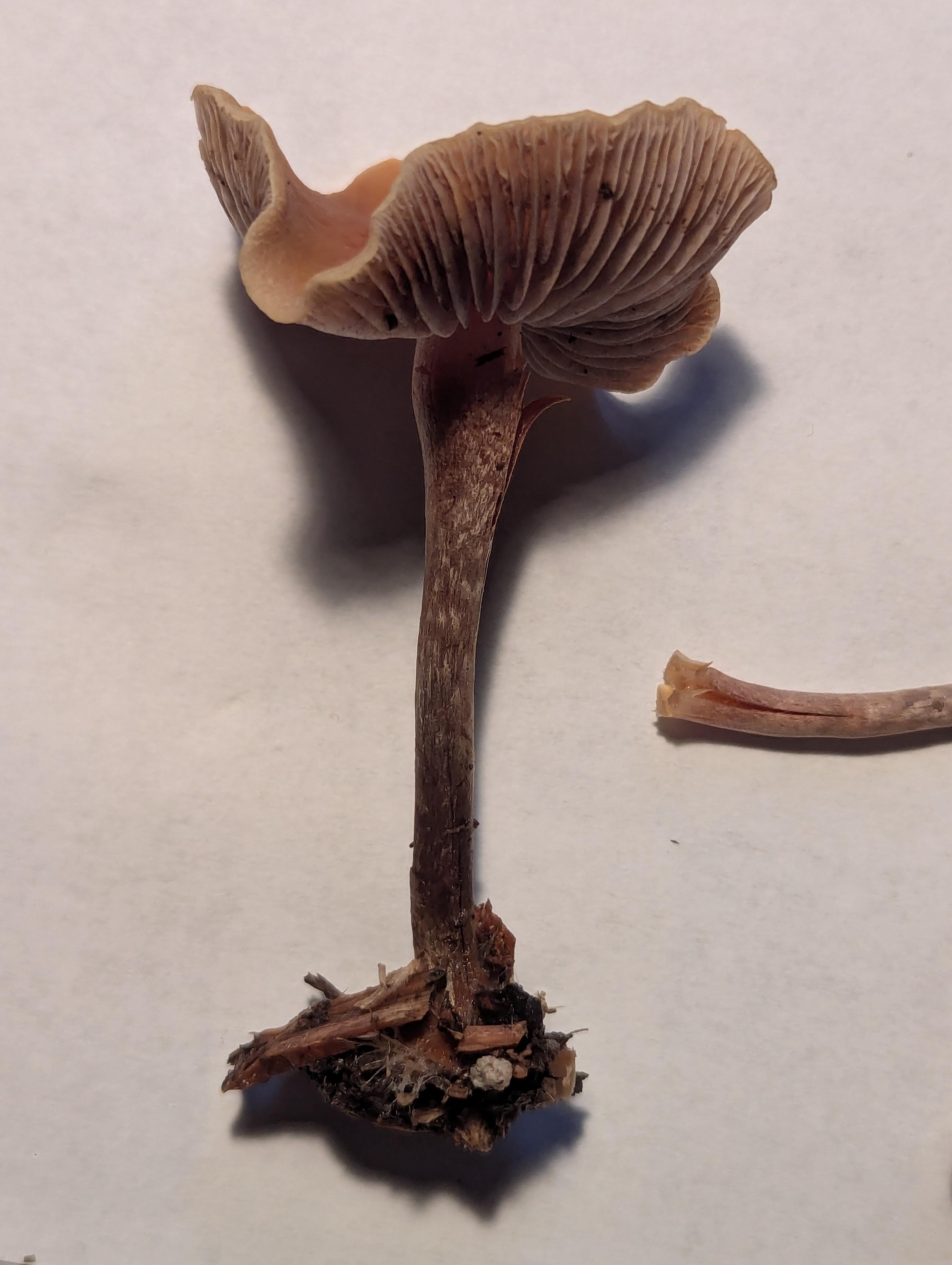
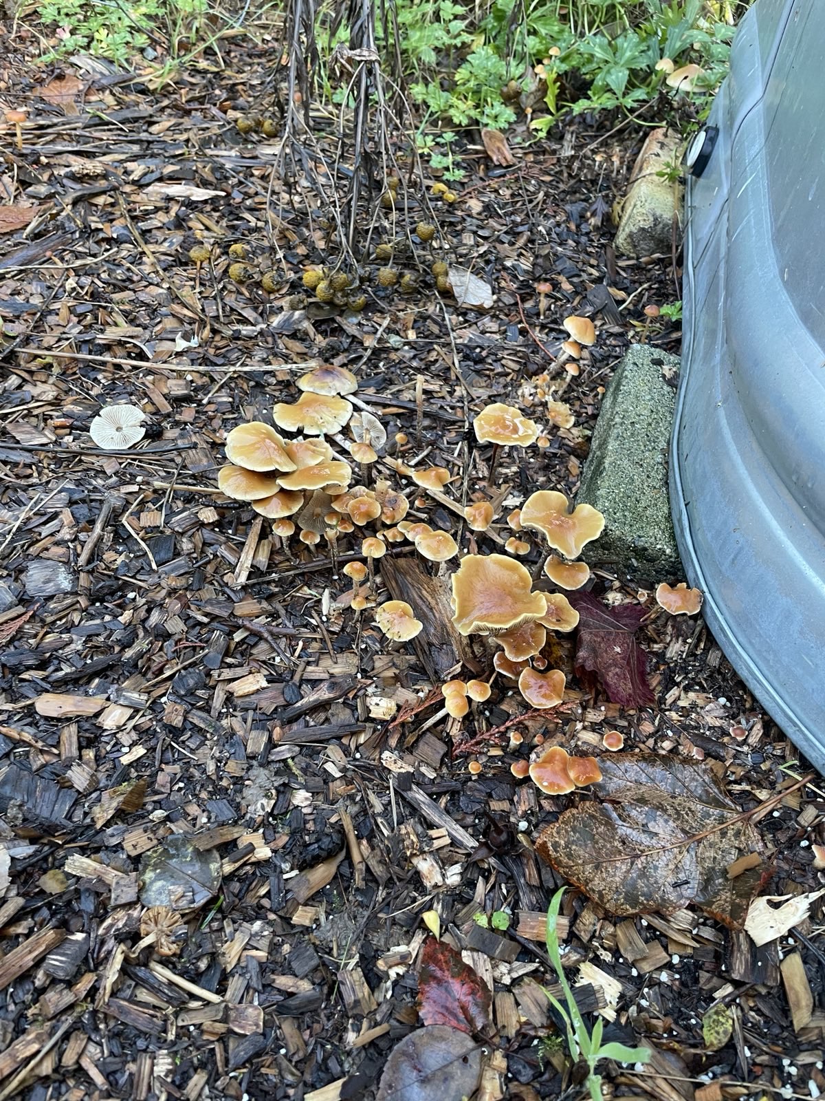
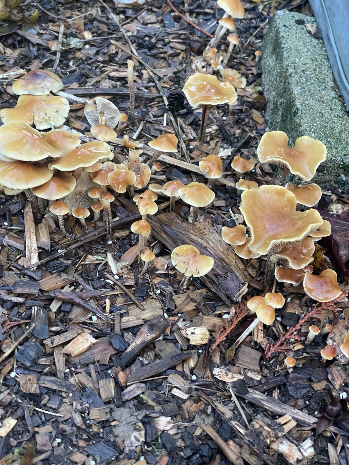
They were found in mid-november in the Salish Coast region of Cascadia. They were growing out of woodchips composed of a mixture of western hemlock (majority), and western red cedar.

Side view of one full mature specimen:

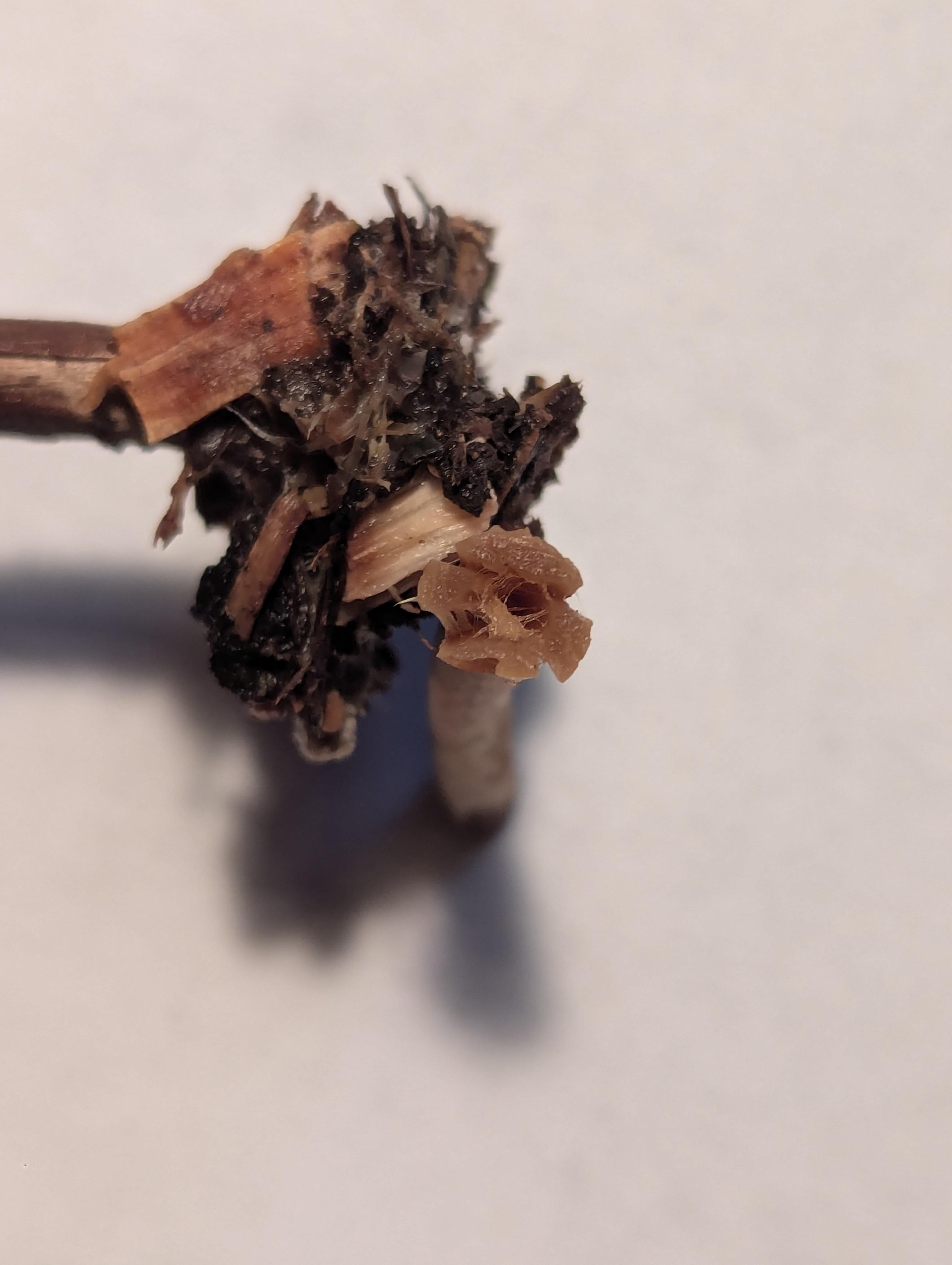
A group with a sample of the substrate (the cap appears to be umbonate):

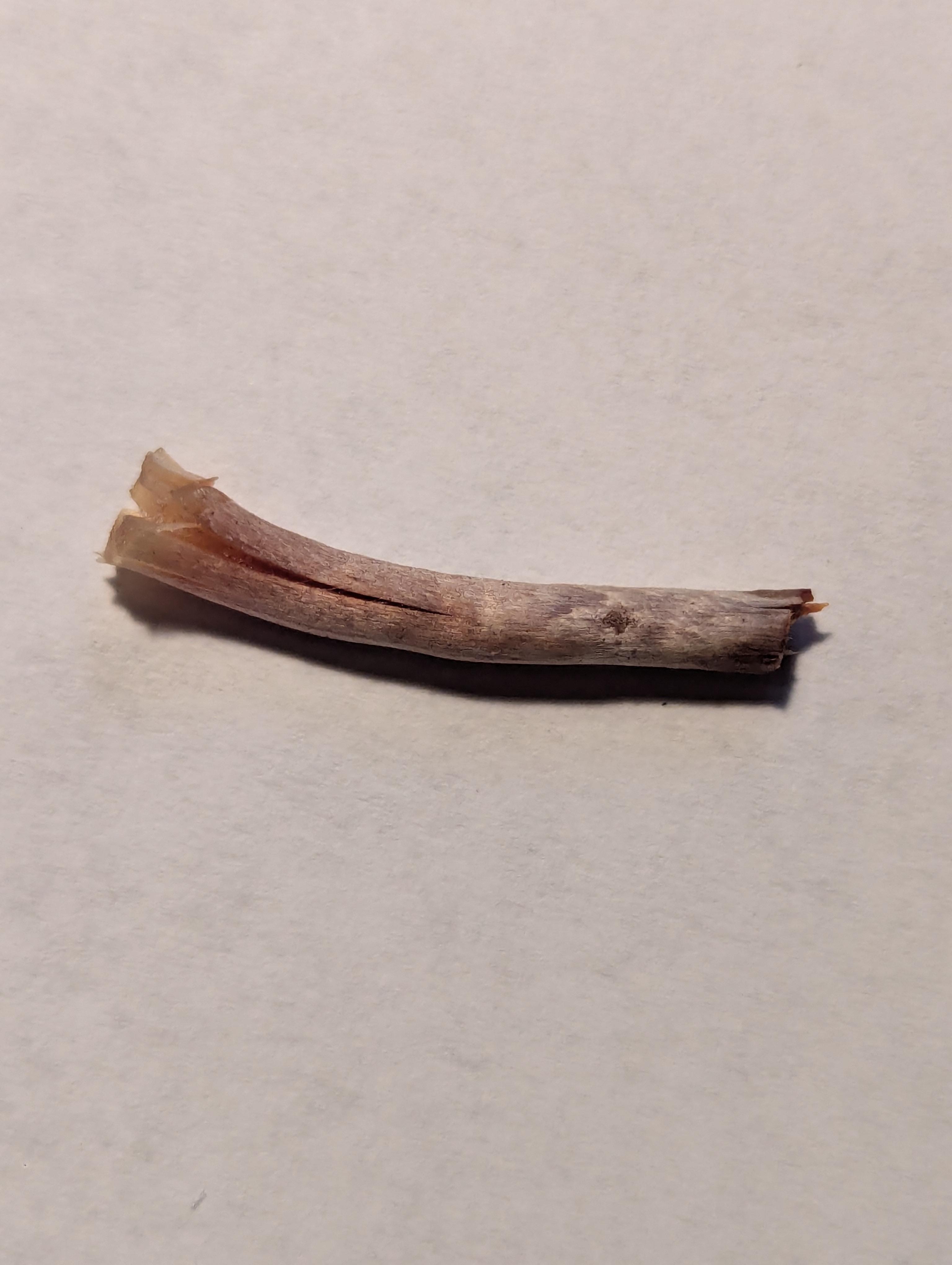
A closeup side view, and internal view of the stem (it appears to be hollow):

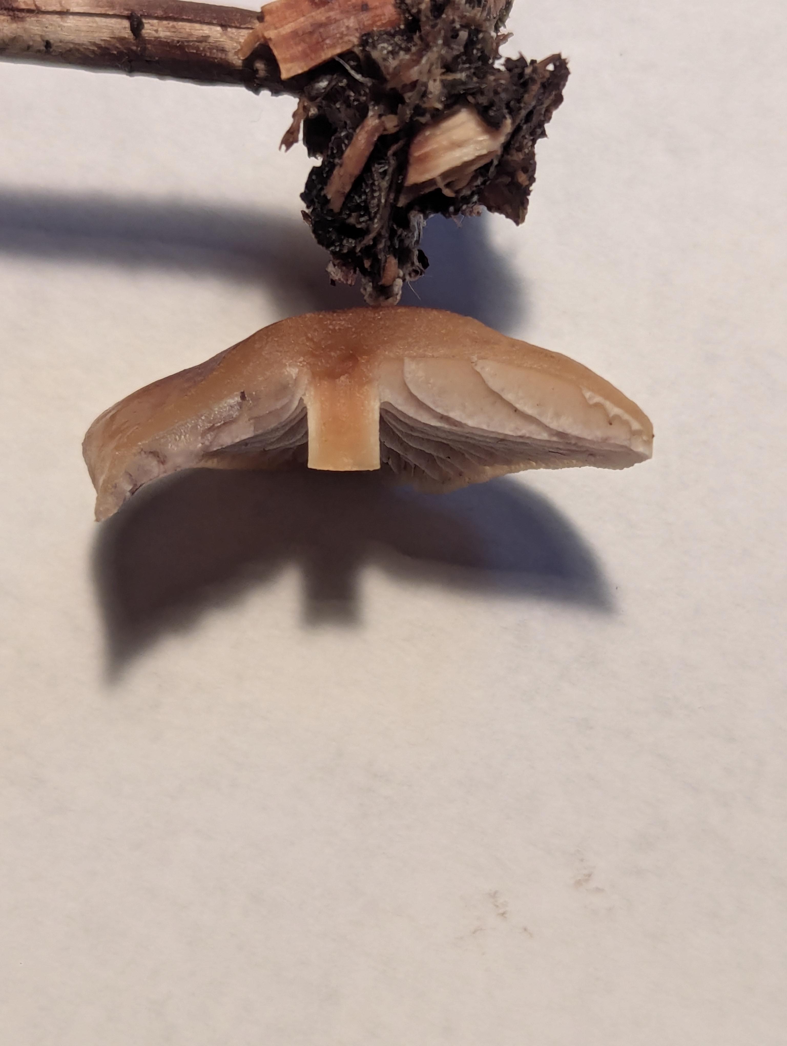
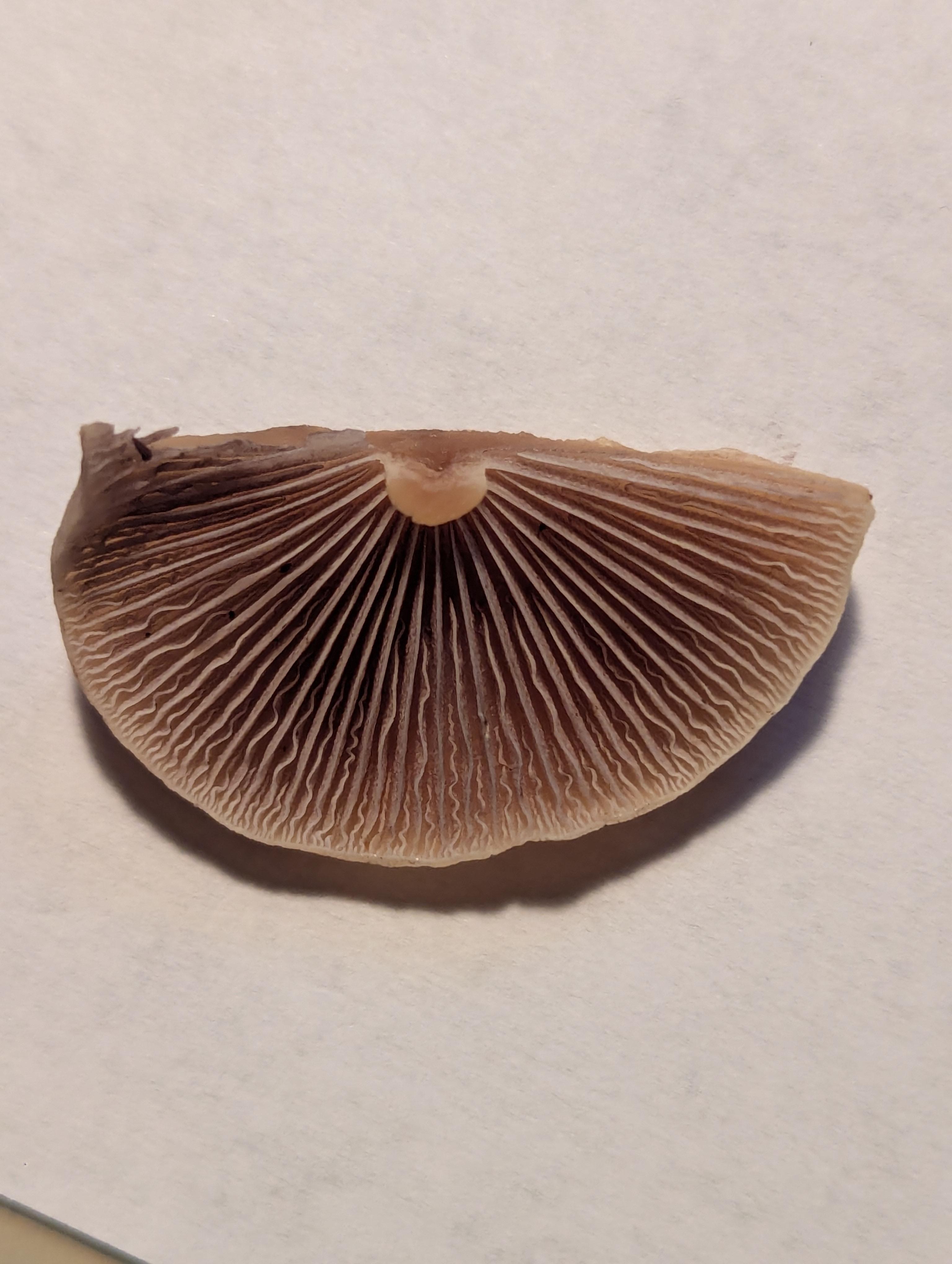
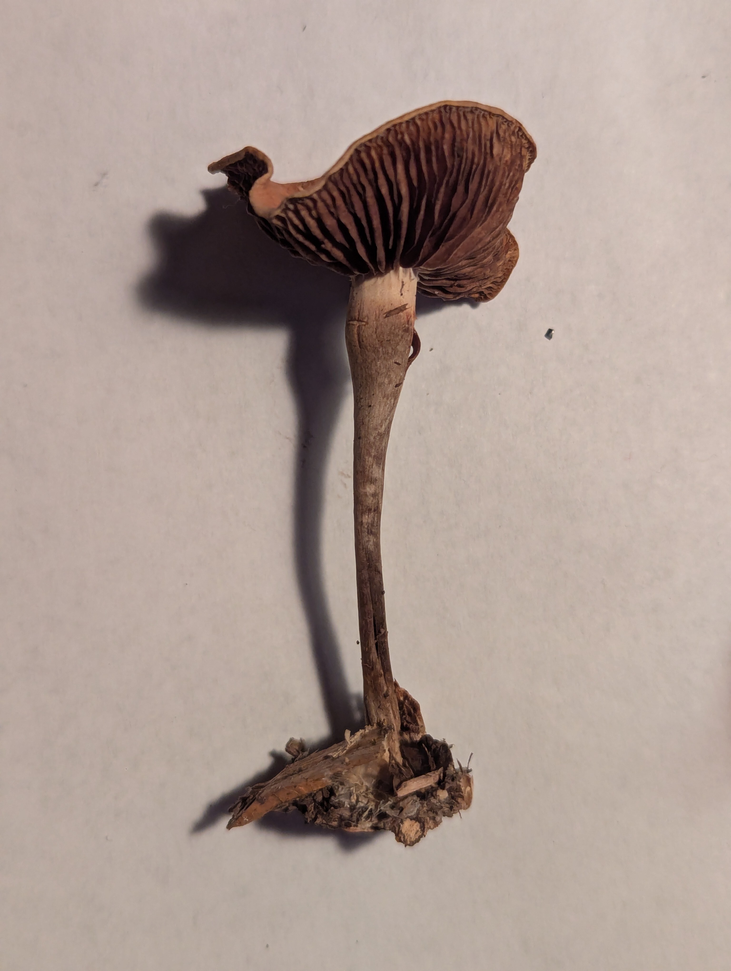
Cross section of the gills โ€” they appear to be adnate, or sub-decurrent:

Underside of view of the gills:

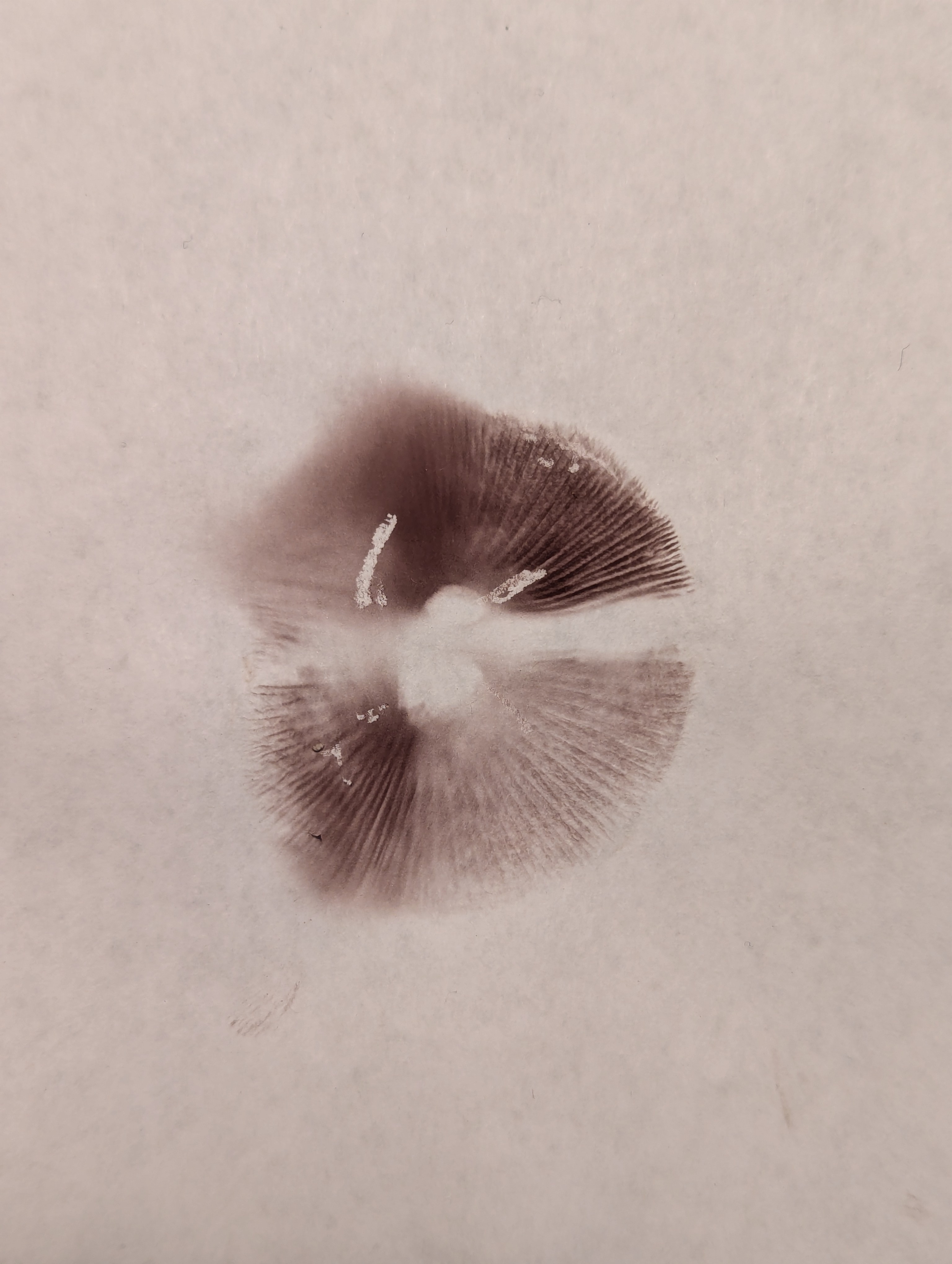
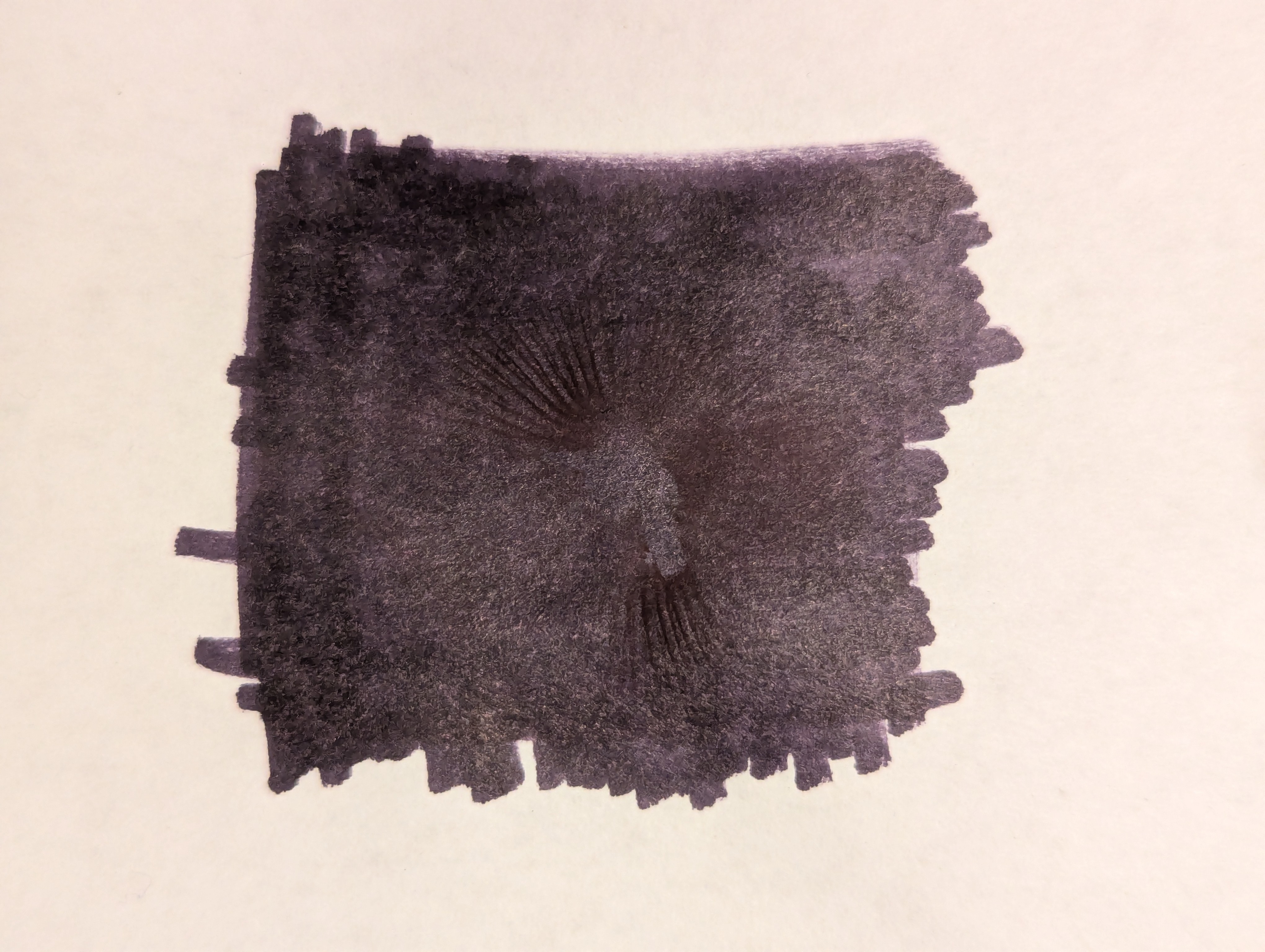
Spore print (first on white background (the split is due to two halves), second on a black background):

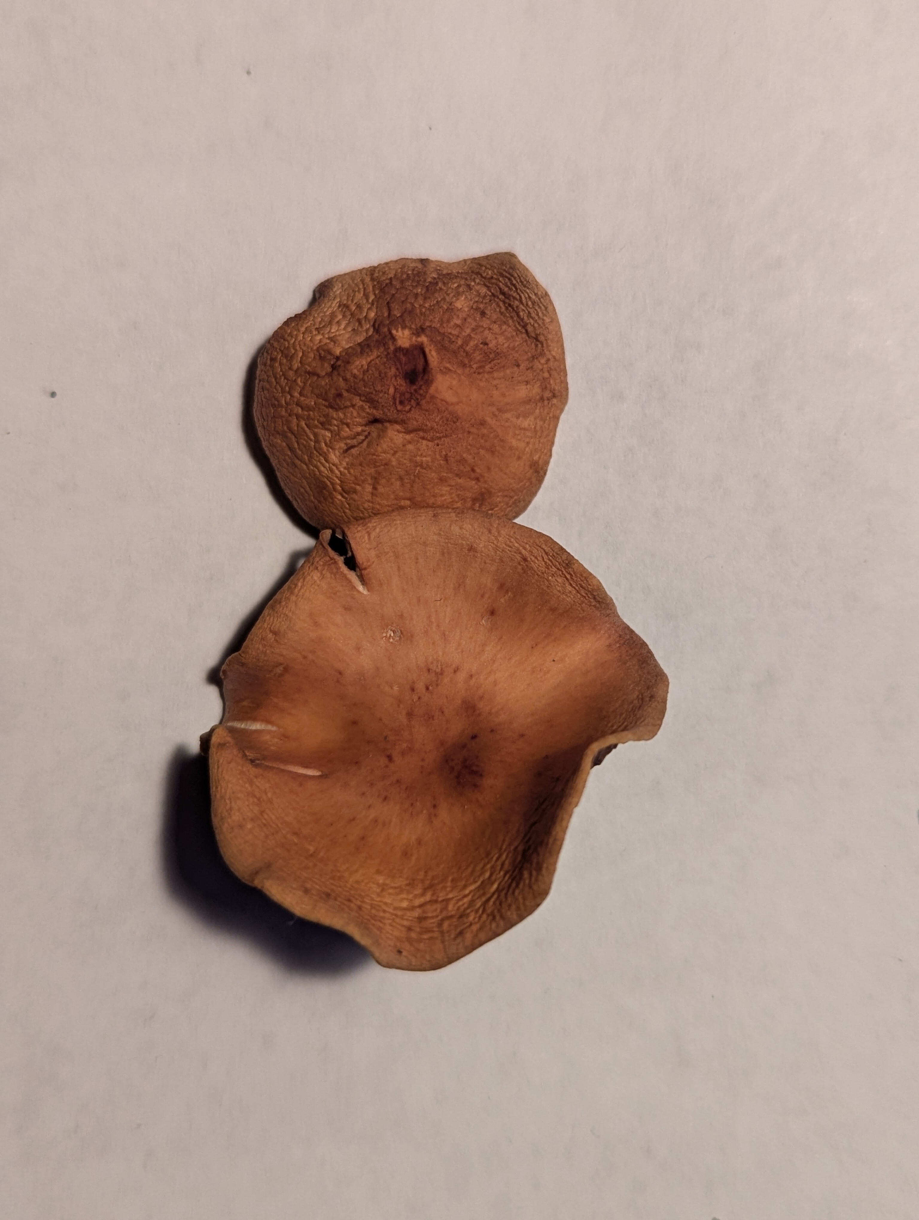
Examples specimens once dried:

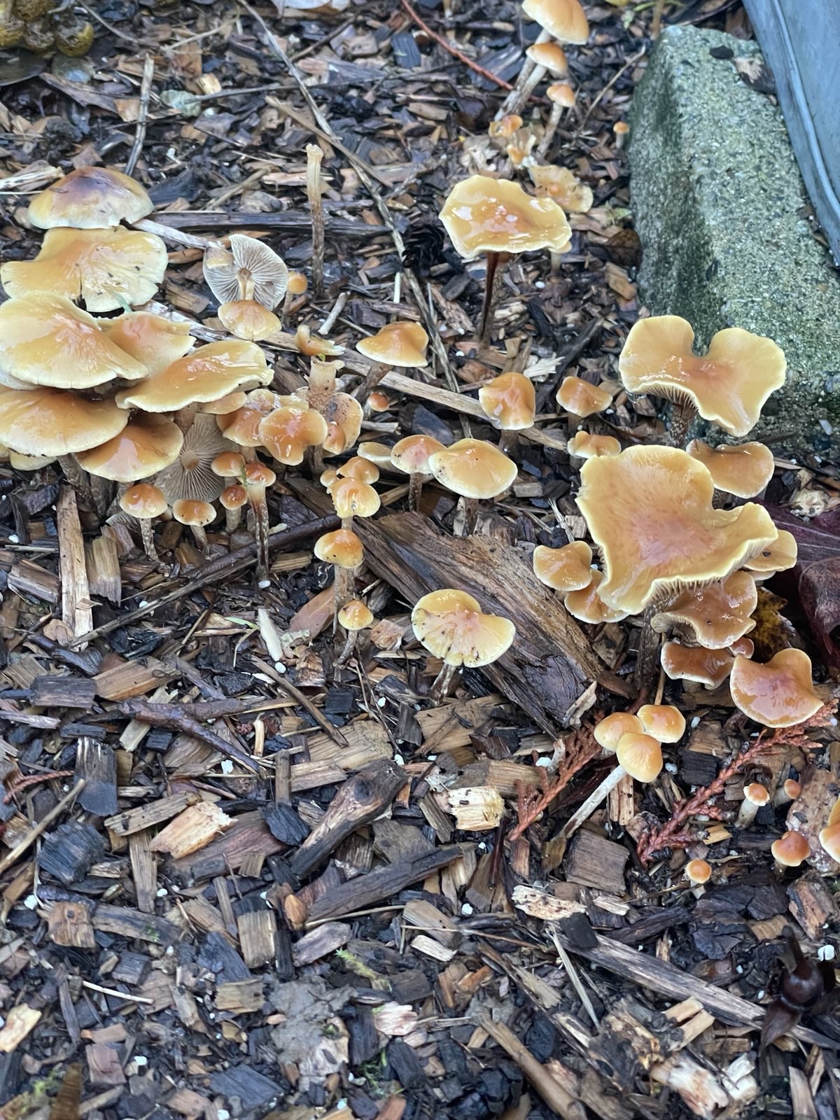
Examples of the colony, and the location/substrate in which it was growing:


Cross-posts:

you are viewing a single comment's thread
view the rest of the comments
[โ€“] [email protected] 1 points 1 month ago

Maybe you could optimise the spore print picture.

I personally like to take the prints on aluminium foil, because

  1. It gives okay contrast for both bright and dark spores.
  2. It's cheap
  3. It's almost sterile and can easily be disinfected
  4. You can store the prints better by folding the foil.
  5. You can ship and share the prints way easier without risks (microscopy slides breaking, paper soaking up, etc.)

Neat Idea to use aluminum foil! I'll have to try aluminum foil next time. I also hadn't considered physically storing the prints for future reference ยญโ€” it likely would make sense to do so.