this post was submitted on 03 Aug 2023
62 points (100.0% liked)
Radiology
594 readers
1 users here now
A community for all things related to medical imaging!
RULES:
-1. Please follow the Lemmy.World Server Rules.
-1A. Please be civil.
-1B. Please be respectful when discussing medical cases.
While we do not wish to impose a somber tone upon this community, please remember that there are real patients behind the images.
-2. No patient-identifiable information. There is zero tolerance for breaching patient confidentiality laws. De-identified information is allowed under HIPAA, the US patient confidentiality law. Consent is not required when posting de-identified information.
-3. No requests for medical advice or second opinions. Please go to your physician/provider for assistance. Online strangers will never know your clinical history as well as your actual healthcare team, nor will the images posted here be of sufficient quality or completeness for diagnostic interpretation. Any content or discussions in this Community should be considered for educational purposes only, and their accuracy or quality with regards to standard of care cannot be guaranteed.
-4. No spam or advertising. Products or companies that are mentioned as a natural course of discussion are allowed.
-5. Please do not spread misinformation.
-6. Moderators have final say in their decisions. Please, no rules lawyering.
Posts:
Only moderators may post in this community pending Lemmy's implementation of a moderator-approval process. Until that happens, you may post general comments or questions in the stickied megathread. You may also request the moderators to post on your behalf via DM, pending time and availability.
founded 1 year ago
MODERATORS
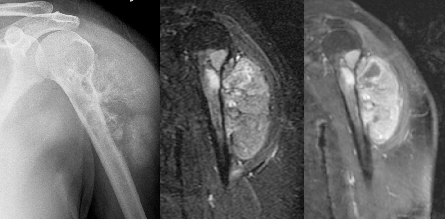
I find these posts fascinating, but I am a layperson with only whatever medical knowledge a doctor father who loved to share imparted. I can somewhat understand these images, and the MRI looks obviously anomalous to me, but I always struggle with X-rays! Would anyone care to help me understand what exactly it is in the X-ray that indicates an issue? To my uneducated eyes, it doesn’t seem that different from a healthy shoulder.
Is it the fact that there’s “cloudiness” between the bone and skin? I guess I can somewhat pick that out now, but I just don’t feel confident I would ever have noticed it on my own without the context of this post.
Hi. Try to match the outline of the humerus to that on the radiograph. Then the big bright mass to the right of the humerus (going by the picture) is the cancer. Now see if you can make out the rough outline of it on the radiograph.
The cloudiness you're seeing is calcification within the tumor. Specifically, the outward "tendrils" (as someone else called it) exploding out from the margin of the humerus is really aggressive periosteal reaction, which I've posted a few examples before already: one, two.
I think I understand that, thank you. Your other examples are very helpful, though it took some study before I could tell the differences. Would I be right in saying that all tumors calcify, and that the level of calcification (as well as the orientation of the “tendrils”) indicates the progression of the tumor?
No they don't all calcify.
The tumor here is growing from the bone and destroying it. The normal bone cells are trying to repair by making more bone. If faster the tumor destroys and grows, the less smooth the repaired bone can look.