Radiology

598 readers
1 users here now

A community for all things related to medical imaging!

RULES:

-1. Please follow the Lemmy.World Server Rules.

-1A. Please be civil.

-1B. Please be respectful when discussing medical cases. While we do not wish to impose a somber tone upon this community, please remember that there are real patients behind the images.

-2. No patient-identifiable information. There is zero tolerance for breaching patient confidentiality laws. De-identified information is allowed under HIPAA, the US patient confidentiality law. Consent is not required when posting de-identified information.

-3. No requests for medical advice or second opinions. Please go to your physician/provider for assistance. Online strangers will never know your clinical history as well as your actual healthcare team, nor will the images posted here be of sufficient quality or completeness for diagnostic interpretation. Any content or discussions in this Community should be considered for educational purposes only, and their accuracy or quality with regards to standard of care cannot be guaranteed.

-4. No spam or advertising. Products or companies that are mentioned as a natural course of discussion are allowed.

-5. Please do not spread misinformation.

-6. Moderators have final say in their decisions. Please, no rules lawyering.

Posts:

Only moderators may post in this community pending Lemmy's implementation of a moderator-approval process. Until that happens, you may post general comments or questions in the stickied megathread. You may also request the moderators to post on your behalf via DM, pending time and availability.
founded 2 years ago
MODERATORS
51


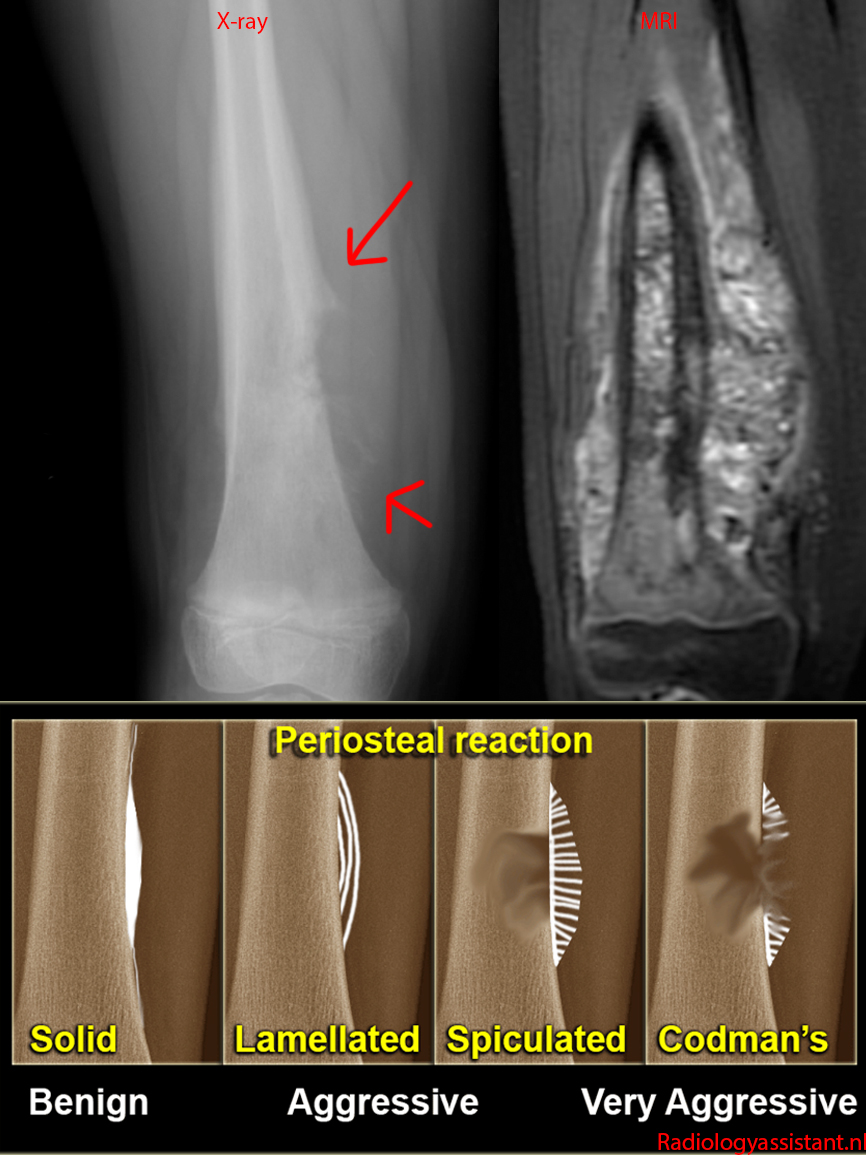
7 year old with conventional (intramedullary high-grade) osteosarcoma of the femur.

X-ray shows an aggressive bone lesion with Codman's triangles and extension into the adjacent soft tissues.

MRI shows the full extent of the tumor much better - it is involving the full thickness of the femur, has much further soft tissue extension than suggested by the Codman's triangles, and even extends into the soft tissues on the other side.

52
2
submitted 2 years ago* (last edited 2 years ago) by Spectator to c/radiology


68 year old with several months of worsening fatigue and 20 pound weight loss. Physical exam notable for a firm, nodular liver.

CT [top images]: Large round, heterogeneous mass, this time in the right hepatic lobe. See here for a left hepatic cholangio. This was initially thought to be a large hemangioma.

MR [bottom left image - T2 FS, bottom right image - postcontrast LAVA]: Large, very heterogeneous mass, partially compressing the inferior vena cava.

This mass was also not survivable.

53


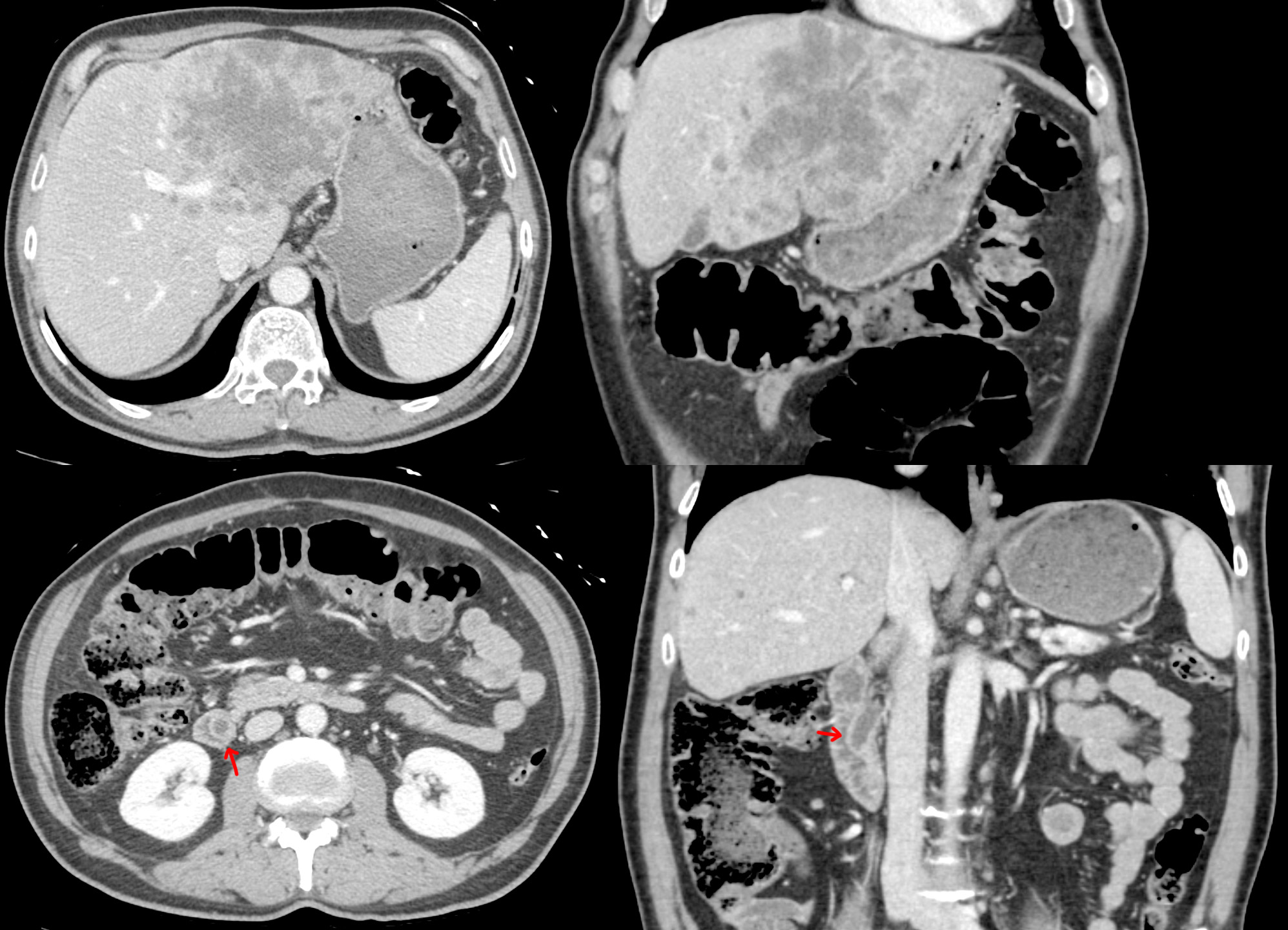
59 year old with 6 months of abdominal pain and weight loss. CA 19-9 tumor marker measured 92,000. CEA tumor marker measured 205.

CT [top images]: Large heterogeneous mass with irregular borders involving the left hepatic lobe.

CT [bottom images]: Protrusion of the distal common bile duct (red arrow) into the duodenum, likely represent a Todani type III bile cyst / choledochocele.

This aggressive cancer was not survivable.

54


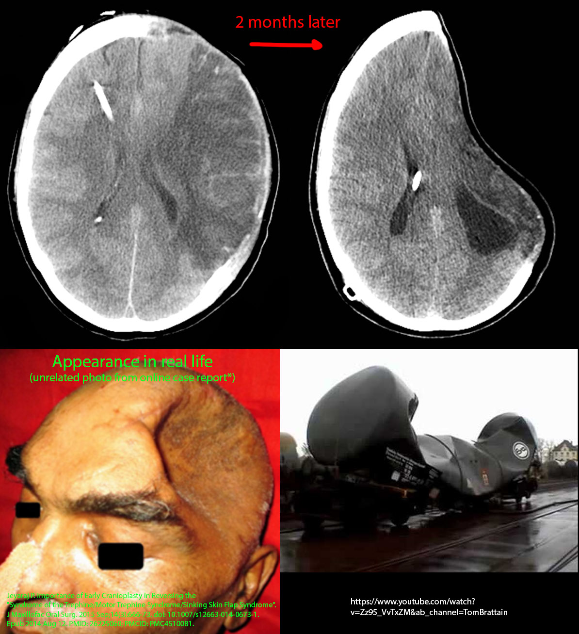
The recent event with the Titan submersible reminded me of this case.

33 year old female with a ruptured aneurysm that ultimately caused a large left-sided stroke with massive brain swelling. This necessitated a decompressive craniectomy to release the pressure and placement of a ventricular shunt.

2 months later, she presented with a sunken appearance where the craniectomy was done and new seizures.

CT shows new sunken appearance of the brain at the craniectomy site and a paradoxical, left-to-right midline shift away from the craniectomy.

Sunken skin flap syndrome (also known as trephine syndrome) is caused by atmospheric pressure pushing against the brain, with risk factor being ventricular shunt or drain (which will reduce intracranial pressure). It is a known complication of craniectomies, and the treatment is cranioplasty (place the missing skull back on). She had already been planned to get the cranioplasty, but this problem developed a little earlier than anticipated. Note: a depressed appearance by itself is not a major issue (aside from cosmetic) - it is the presence of seizure or other neurologic symptoms that necessitate more urgent management.

55


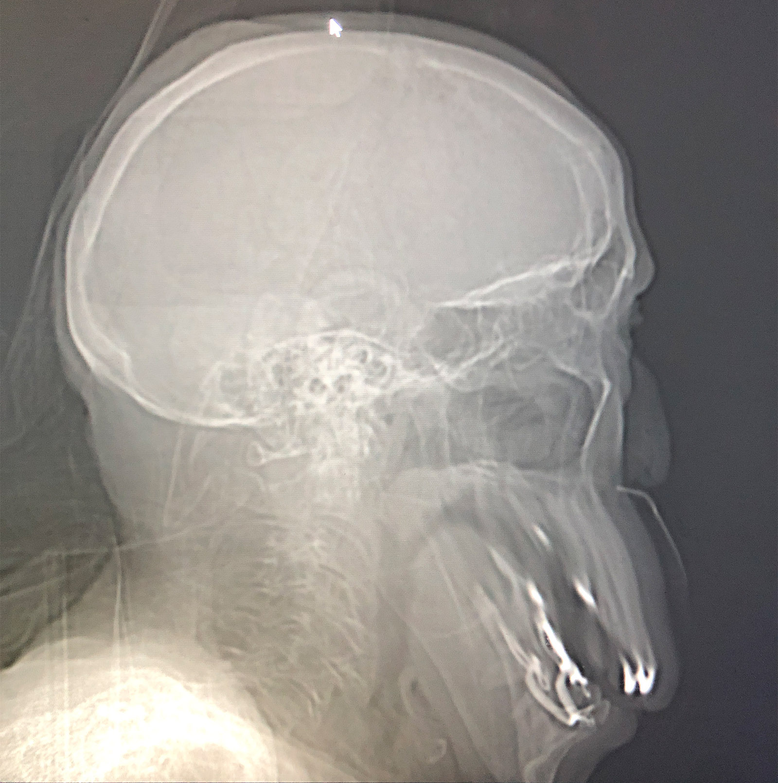
The patient moved his jaw while being scanned.

56


8 year old with a history of trisomy 21 (Down syndrome) with tachycardia.

Chest x-ray shows prominent interstitial markings and cystic changes throughout the lungs. An atrial septal defect occlusion device is present.

CTs show prominent interstitial markings and ground glass opacities as well as numerous cysts that are predominantly subpleural (peripheral) in location.

Cystic subpleural changes are a known finding of trisomy 21, although with a relatively limited number of reports in the literature. This patient likely also has additional chronic lung disease based on the presence of the interstitial markings and ground glass opacities that may be related to prematurity or prior infectious/inflammatory processes.

57


CT obtained for some unrelated symptoms with an incidental finding.

Near the top, the two kidneys looked normal* [top image]. Near the bottom, the two kidneys wanted to join up into one kidney [bottom image].

*Technically, a little bit of renal malrotation due to the abnormal positioning from the horseshoe morphology.

This is generally a benign finding, but the abnormal shape might contribute to poor urinary drainage, which can cause renal stones, urinary infection, increased risk for cancer from the chronic inflammation caused by repeated infections, or increased risk of injury from trauma.

58
1
submitted 2 years ago* (last edited 2 years ago) by Spectator to c/radiology


26 year old female with 2-3 months of abdominal enlargement and weight gain.

CT shows a -massive- multicystic mass filling the entire abdominal cavity and displacing the normal organs. The mass extends from the pelvis to above the belly button, ~25-30 cm in length.

The patient underwent exploratory laparotomy. The mass originated from the right ovary, which was resected. The omentum, regional lymph nodes, and appendix were also removed. Luckily, there was no recurrence of the tumor at last follow-up 2 years later, and she went on to have kids.

59


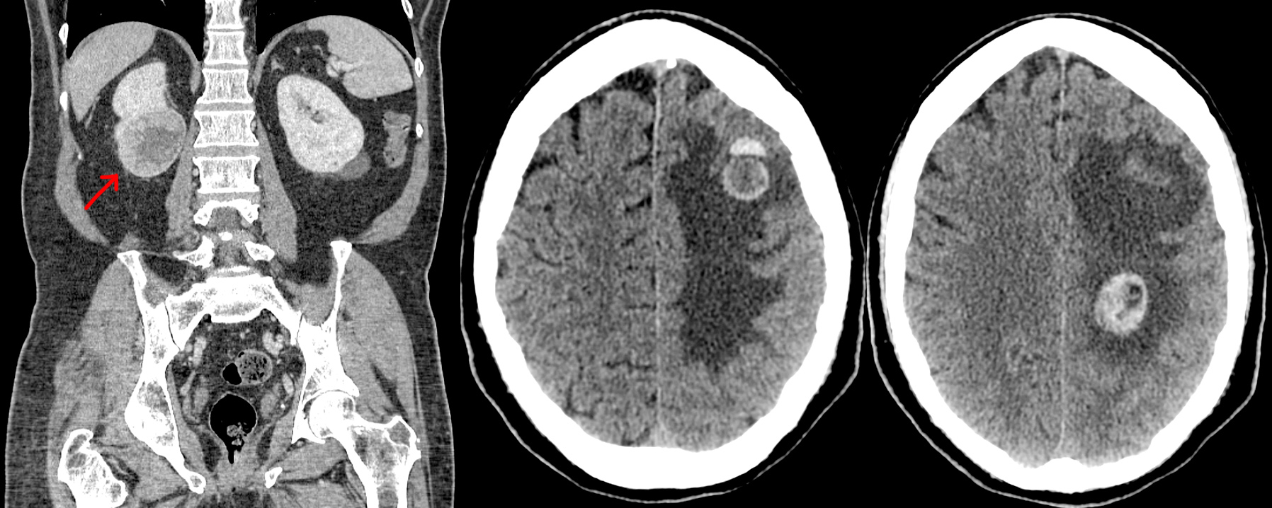
61 year old male, who came in with symptoms of right arm weakness and 3 months of weight loss despite trying to gain weight.

Head CT shows multiple round lesions in the (patient's) left cerebral hemisphere (right side on images), which likely account for the right-sided weakness. These lesions show hemorrhage as well as substantial surrounding brain edema.

A work-up for metastatic disease included a CT of the abdomen/pelvis, which showed the primary mass in the right kidney.

60


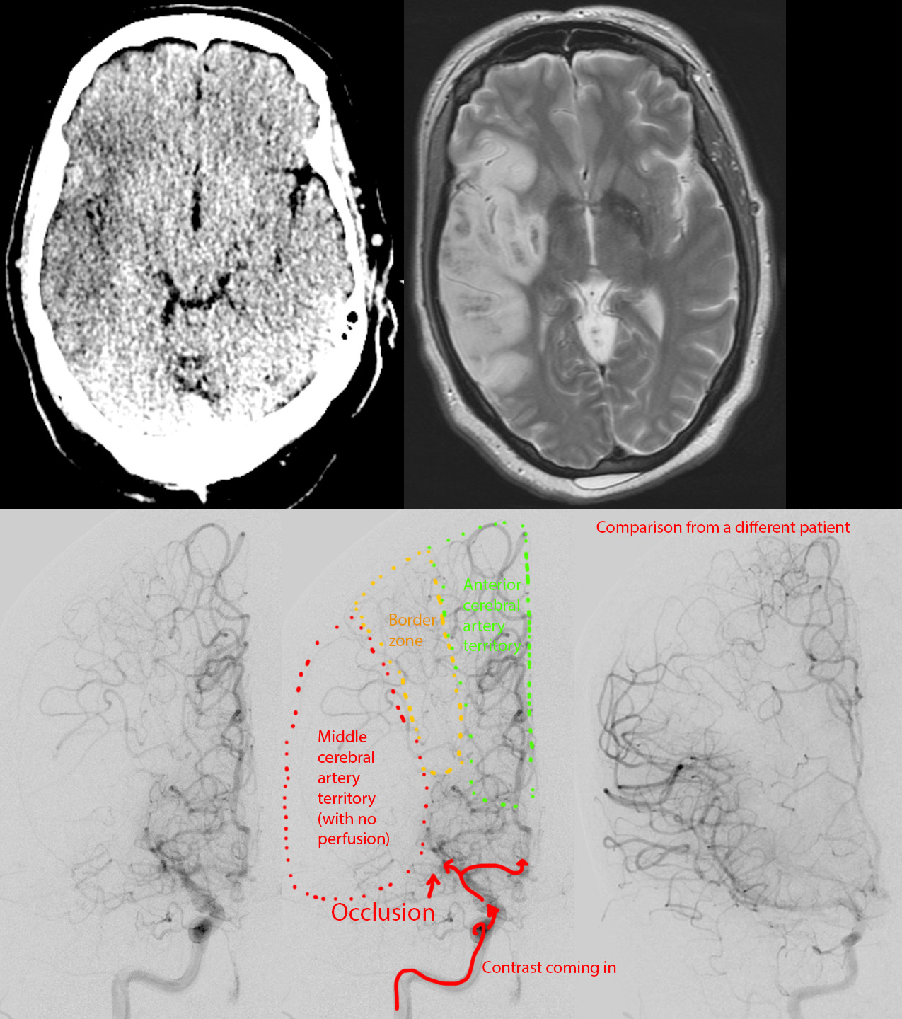
Female in her 50s, with a history of meth abuse. She suddenly developed confusion and left-sided weakness while talking on the phone.

Dual energy CT virtual noncontrast [top left image] shows a large rectangular area of hypodensity in the right middle cerebral artery territory.

Cerebral angiogram shows occlusion of the right MCA with no filling of its distal branches or the brain parenchyma [bottom left, bottom middle]. A stent retriever mechanical thrombectomy was performed [not shown].

MRI done 1 day later shows the infarct with the affected area showing brain swelling [top right].

61


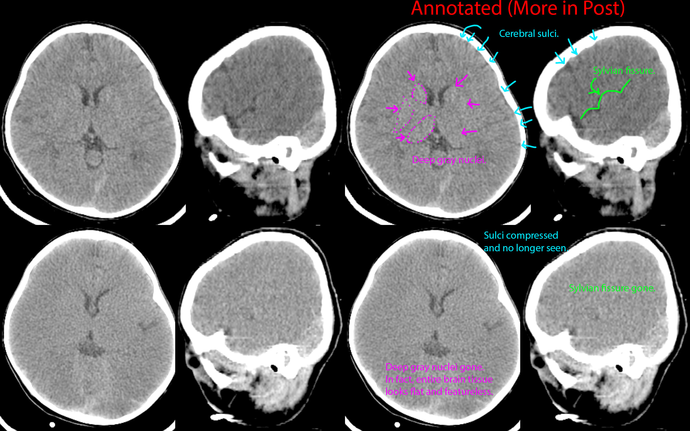
This patient had a complicated medical history including severe congenital heart disease. He had cardiac arrest requiring one hour of resuscitation.

He never regained consciousness, and further evaluation with EEG one day later revealed minimal brain activity consistent with severe dysfunction; a CT of the head was done that did not show any significant findings at that time [top images].

Two days after that, repeat EEG and head CT were done due to no improvement in neurologic status. EEG showed no activity, and CT showed developing signs of diffuse cerebral edema and anoxic brain injury. After discussing goals of care with patient's family, support was withdrawn, and the patient passed away.

62
63


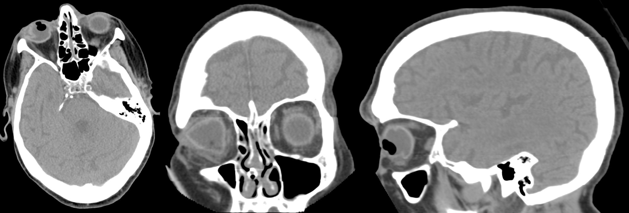
Past medical history of seizure. Had an unwitnessed fall.

CT shows right globe with abnormal irregular morphology, abnormal location/appearance of the lens, and presence of air within the globe. On the coronal view, there is fluid infiltrating the surrounding orbital fat. A left forehead hematoma is also present.

64


Otherwise healthy newborn with "abnormal back finding."

Ultrasound shows, underneath the back "mass," a defect of the posterior spine through which the spinal sac protrudes.

MRI confirms the defect and spinal sac protrusion. Additionally, there was some tissue that was thought to represent neural placode within the herniated sac. There was no obvious fat-containing mass within to suggest dermoid by imaging.

Surgery showed absent T6-T7 spinal lamina with a fibrous tract extending from the back of the spinal cord to the skin surface. Hair and other skin-related debris extruded from the tract as it was opened. The entire fibrous tract and its contents were removed.

65


If you're planning on getting an MRI, avoid wearing Lululemon, especially their silver-impregnated stuff - the strong changing magnetic fields can cause the clothing to heat up to the point of causing burns (just Google for news articles).

66


68 year old with nausea and vomiting.

CT shows very thickened irregular walls at the gastric outlet (arrows) with distended stomach.

Biopsy showed poorly-differentiated adenocarcinoma with signet ring cells.

67


12 year old with pectus deformity causing fatigue and chest pain. CT shows the really deep sternum indenting the heart. Cardiac echo (not shown) reported right ventricular compression to the point it was affecting systolic function. The patient had Nuss bars placed to correct the deformity.

68


On the left side, 2 x-rays of 2 different patients with 2 different objects in their pelvis. Internet denizens would no doubt instantly diagnose these as accidental rectal foreign bodies from people falling on their butts. However, these are both pessaries used to treat pelvic organ prolapse. The top one is an inflatoball pessary. The bottom one is a ring-shaped pessary that we see on edge.

69


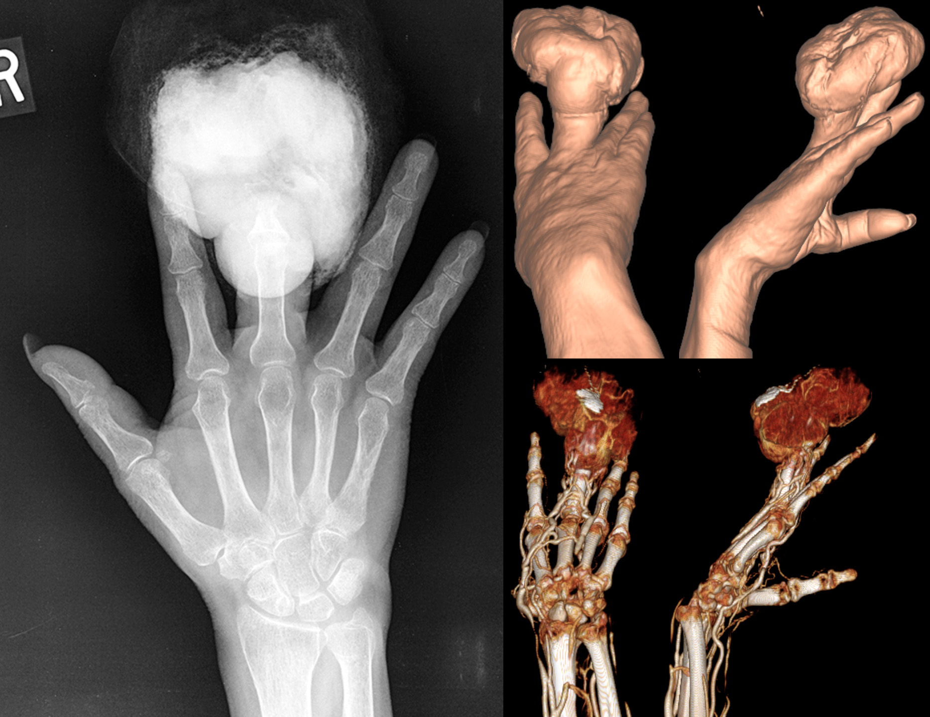
54 year old female with a history of minor injury to the finger 6 months before these studies were obtained, subsequently developed an infection requiring debridement. The wound was then stable until 2 months before, when a large, fungating, hemorrhagic mass grew.

X-ray shows large radiopaque mass eroding the distal finger (distal phalanx and distal portion of the middle phalanx).

CT 3D surface reconstructions show the morphology of the mass.

CTA 3D reconstruction shows the mass is very hypervascular.

Lymph node scintigraphy was performed showing a sentinel node at the axilla (not shown). The patient underwent amputation of the finger with axillary sentinel node biopsy, which was positive for metastatic melanoma. The patient was then lost to follow-up.

70


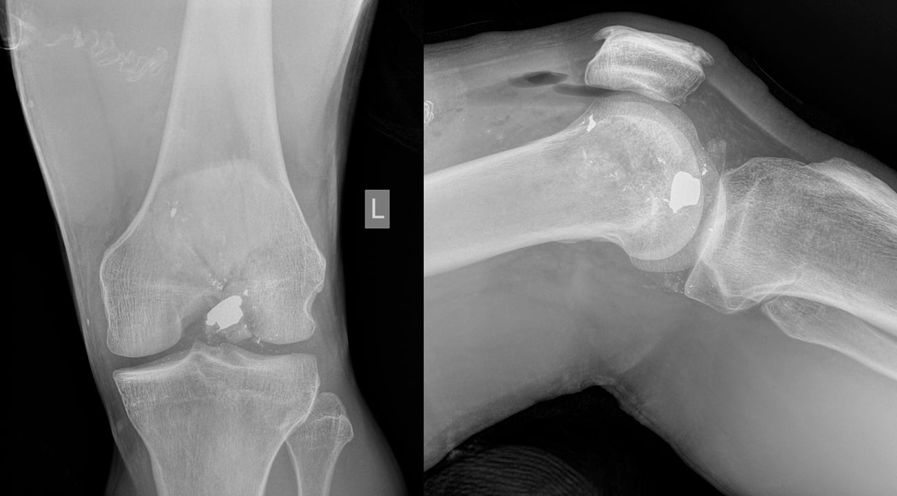
38 year old guy fired his 45 mm into his knee while cleaning the gun. No exit wound.

There is a comminuted fracture of the distal femur with multiple bullet fragments, including the largest at the intercondylar notch. There is gas/air in the knee joint, forming air-fluid levels on lateral view. There are additional foci of gas in the surrounding soft tissues.

Patient underwent arthrotomy for foreign body removal and fixation of the supracondylar/intercondylar femoral fracture.

71


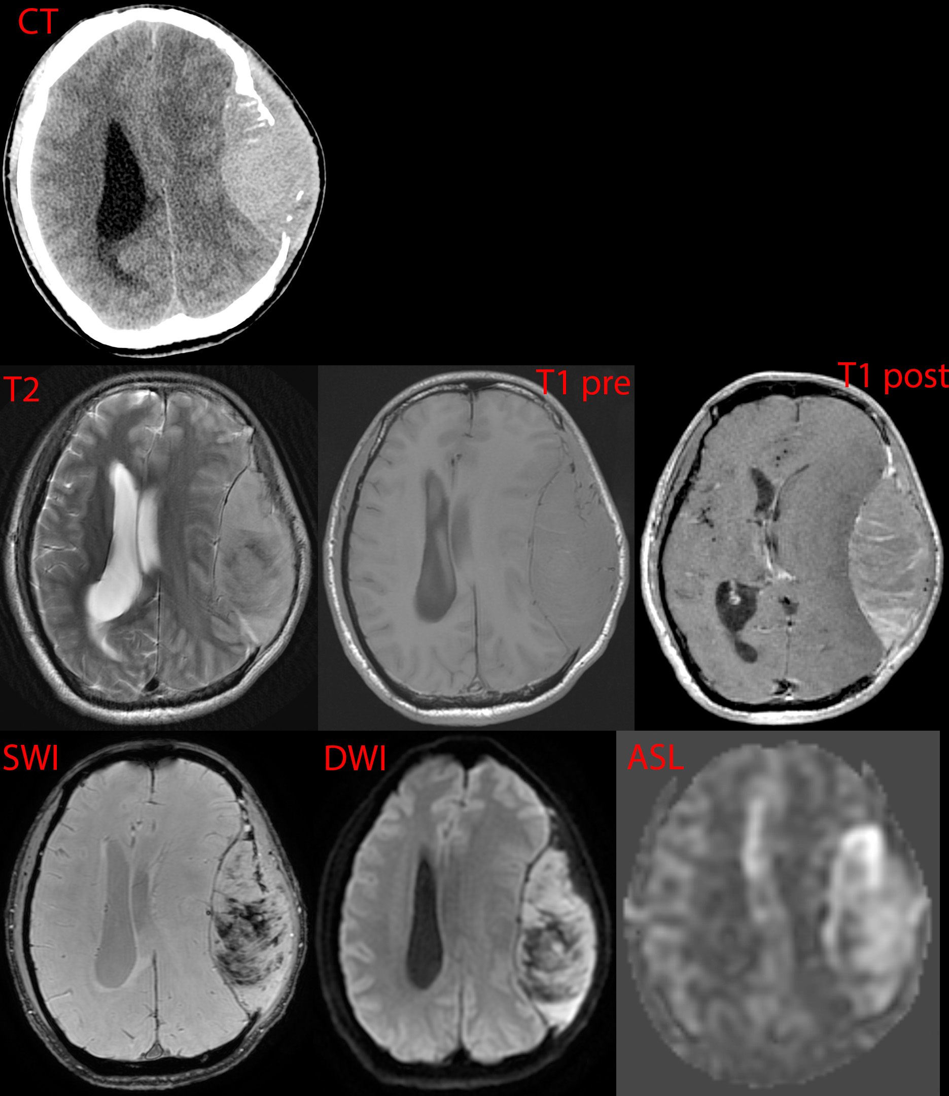
41 year old male with history of HIV-AIDS, meth-abuse, presents with progressive headache and confusion over days.

CT shows a large mass destroying the left frontoparietal calvarium and causing significant mass effect on the underlying brain.

MR shows a heterogeneous enhancing mass with hemorrhage (SWI - dark areas), hypercellularity (DWI - mildly bright areas), and hyperperfusion (ASL - bright areas), features suspicious for high-grade tumor.

72


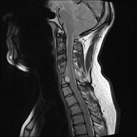
This is my neck. I was born with this and it was discovered when I was 41. There are fused vertebrae from C4-C7, malformed discs, deformed vertebrae, a bulging disc touching my spinal cord and the dermoid tumour on my spinal cord.

73


62 year old female with a history of tobacco, opioid, and meth abuse as well as heart attack requiring coronary bypass and stents. She presents with bilateral lower extremity claudication (painful fatigue with physical activity) starting from her glutes.

CT angiogram shows occlusion of the lower aorta (red markings) as well as both common iliac arteries.

Vascular ultrasound shows very poor arterial waveforms throughout the lower extremities, essentially flat/absent below the knee.

She underwent a transabdominal aortobifemoral bypass to treat this.

Radiopedia article on Leriche syndrome.

Meth is bad.

74


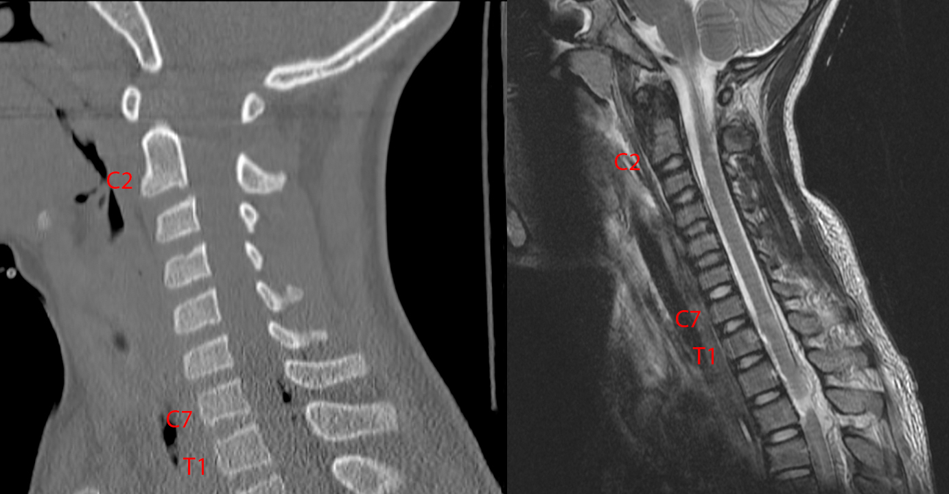
Very unfortunate case of a young patient, who was crossing the street on a bike when he was struck by an SUV. The patient had brief loss of consciousness with decorticate posturing. Following that, he had reported no sensation and total weakness from the waist down, with physical exam showing total sensory and motor loss from the nipple level (T4) and below.

CT shows no fractures but some abnormal amount of gas/air in the back of the spinal canal at C7-T1.

MR shows complete transection of the spinal cord with 0.5 cm gap at T3 - goes well with the physical exam finding. There is a dorsal epidural hematoma. There was also disruption of the anterior longitudinal ligament, posterior longitudinal ligament, and ligamentum flavum at T3 (not shown).

What probably happened was that instead of a fracture, the traumatic force went through the ligaments maintaining spinal alignment, causing the spine at the T3 level to dislocate and shear, cutting through the spinal cord. By the time imaging was done, the bones relocated to their normal position, but the damage had been done.

75


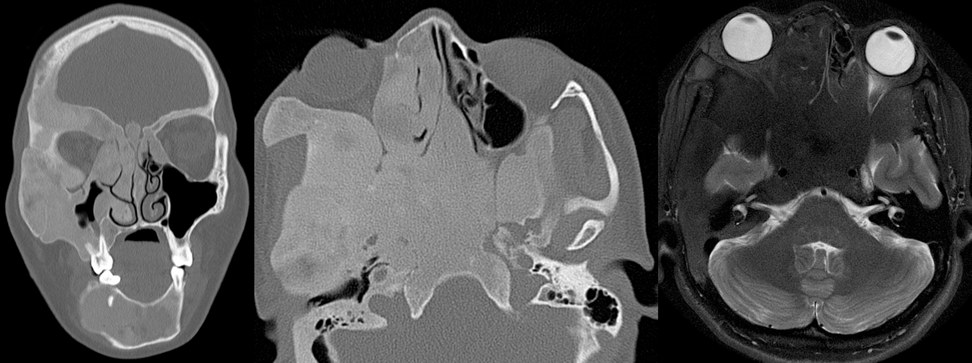
Female in her 20s with (all right-sided) proptosis, intermittent vision loss, facial pain, and mid to lower face numbness.

Coronal and axial CT images show a homogenous bony expansile process that results in narrowing of the soft tissue compartments of the face. As a result, the right eye is pushed forward (proptosis; as seen on the MR image), and many of the right-sided (as well as some left-sided) skull base foramina that carry nerve bundles and blood vessels are severely narrowed.

view more: ‹ prev next ›